It has been found that clots from patients with thrombotic disorders were composed by thin fibers with increased fibrin density and rigidity, and decreased clot lysis rate. The fibrin meshwork structure is characterized by fibrin thickness, fiber density, pores size, and rigidity that reflects fibrin FXIIIa cross-linking and clot structure. įibrin network structure depends on the quality and amount of fibrinogen, on thrombin and calcium concentrations, and on ionic strength, among others.
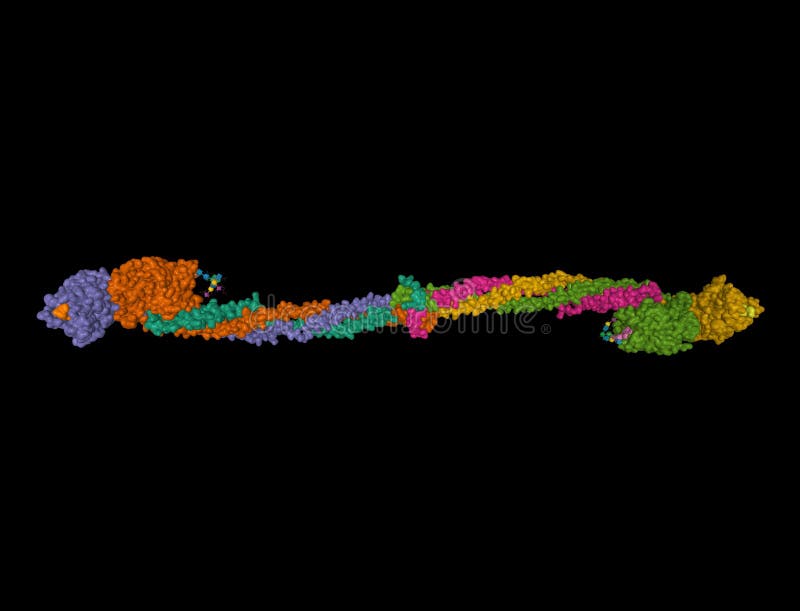
3D MODEL OF FIBRIN JMOL ACTIVATOR
Although tPA is a poor plasminogen activator in solution, at the surface of fibrin the reaction is amplified approximately two orders of magnitude. Furthermore, endothelial cells are involved in fibrinolysis regulation by secreting substances, such as tPA and plasminogen activator inhibitor type 1 (PAI-1). Plasmin is formed from its precursor (plasminogen, Pg) by tissue type plasminogen activator (tPA) at the surface of fibrin. Under physiological conditions, activators, inhibitors, and cofactors finely regulated fibrinolysis. The cross-linked fibrin is deposited in blood vessels and tissues, and plasmin is responsible of the soluble fibrin degradation products. Ĭoagulation and fibrinolysis are activated simultaneously in response to injury. It seems that FpBs release contribute to the lateral aggregation of protofibrils. The lateral aggregation of protofibrils forms the fibrin fibers. The association of fibrin monomers give rise to protofibrils that have two monomer units of width. The release of fibrinopeptides A and B exposes the polymerization sites “A” (knob A) and “B” (knob B), which associate to constitutive polymerization sites located at the C-termini end of the γ chains (“a” or hole a) and β (“b” or hole b). The removal by thrombin of short peptide sequences at Aα A1-R16 (fibrinopeptide A, FpA) and Bβ Q1-R14 (fibrinopeptide B, FpB) triggers the association between the fibrin monomers (fibrinogen molecules devoid of FpA and FpB). įibrinogen does not polymerize spontaneously due to negative charges repulsion at the N-termini of the Aα- and Bβ- chains.
3D MODEL OF FIBRIN JMOL FREE
The C-termini of the Bβ and γ chains are located in the D domain, while that of the Aα chains form a free non well-structured domain, the αC domain.

The fibrinogen molecule consists of two sets of three different polypeptide chains Aα, Bβ, and γ, joined by disulphide bonds at their N-termini E domain. The molecule is 45 nm long and comprises a symmetrical dimer consisting of two outer D domains and a central E domain, linked by α- helical coiled-coil rods. The secretion of fibrinolytic components by HMEC-1 cells seemed to be modulated by integrins that bind RGD ligands.įibrinogen is a 340 kDa plasma glycoprotein that circulates at approximately 2–4 mg/mL. Clot degradation was accentuated at high fibrin concentrations. HMEC-1 cells influence fibrin formation and dissolution as a function of the fibrin content of clots. The secretion of PAI-1 by HMEC-1 cells was unrelated to the presence of fibrin or RGD, 7 × 10 −6 μg/mL per cell and 5 × 10 −6 μg/mL per cell, for the apical (model 1) and basal clots (model 2), respectively.

However, the opposite was found when cells were grown over fibrin: 6 × 10 −6 ng/mL per cell without RGD vs. Secretion of uPA was 13 × 10 −6 ng/mL per cell in the absence of RGD and 8 × 10 −6 ng/mL per cell in the presence of RGD, when clots were formed on the top of HMEC-1.

HMEC-1 retarded dissolution of clots formed onto their surface at 0.5 to 3 mg/mL fibrinogen ( p < 0.05). The rate of fibrin formation increased approximately 1.5-fold at low fibrinogen content (0.5 and 1 mg/mL p < 0.05) compared to the condition without cells however, it was decreased at 2 mg/mL fibrinogen ( p < 0.05) and no differences were found at higher fibrinogen concentrations (3 and 5 mg/mL). The secretion of uPA and PAI-1 by HMEC-1 cells was quantified by ELISA. Clot structure was visualized by laser scanning confocal microscopy (LSCM). Fibrin formation or lysis onto the surface of HMEC-1 cells, was followed by turbidity. Methodsįibrin clots at different fibrinogen concentrations were formed on top of (model 1) or beneath (model 2) the endothelial cells. The aims of the present work were (1) to follow fibrin formation and lysis onto the surface of human dermal microvascular endothelial cells (HMEC-1), and (2) to quantify the secretion of fibrinolytic components in the presence of fibrin. Fibrin provides a temporary matrix at the site of vascular injury.


 0 kommentar(er)
